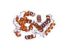
Cyclin

Cyclins are proteins that control the progression of a cell through the cell cycle by activating cyclin-dependent kinases (CDK).[1]
Etymology
[edit]Cyclins were originally discovered by R. Timothy Hunt in 1982 while studying the cell cycle of sea urchins.[2][3]
In an interview for "The Life Scientific" (aired on 13/12/2011) hosted by Jim Al-Khalili, R. Timothy Hunt explained that the name "cyclin" was originally named after his hobby cycling. It was only after the naming did its importance in the cell cycle become apparent. As it was appropriate the name stuck.[4] R. Timothy Hunt: "By the way, the name cyclin, which I coined, was really a joke, it's because I liked cycling so much at the time, but they did come and go in the cell..."[4]
Function
[edit]
Cyclins were originally named because their concentration varies in a cyclical fashion during the cell cycle. (Note that the cyclins are now classified according to their conserved cyclin box structure, and not all these cyclins alter in level through the cell cycle.[5]) The oscillations of the cyclins, namely fluctuations in cyclin gene expression and destruction by the ubiquitin mediated proteasome pathway, induce oscillations in Cdk activity to drive the cell cycle. A cyclin forms a complex with Cdk, which begins to activate, but the complete activation requires phosphorylation as well. Complex formation results in activation of the Cdk active site. Cyclins themselves have no enzymatic activity but have binding sites for some substrates and target the Cdks to specific subcellular locations.[5]
Cyclins, when bound with the dependent kinases, such as the p34/cdc2/cdk1 protein, form the maturation-promoting factor. MPFs activate other proteins through phosphorylation. These phosphorylated proteins, in turn, are responsible for specific events during cell division such as microtubule formation and chromatin remodeling. Cyclins can be divided into four classes based on their behaviour in the cell cycle of vertebrate somatic cells and yeast cells: G1 cyclins, G1/S cyclins, S cyclins, and M cyclins. This division is useful when talking about most cell cycles, but it is not universal as some cyclins have different functions or timing in different cell types.
G1/S Cyclins rise in late G1 and fall in early S phase. The Cdk- G1/S cyclin complex begins to induce the initial processes of DNA replication, primarily by arresting systems that prevent S phase Cdk activity in G1. The cyclins also promote other activities to progress the cell cycle, such as centrosome duplication in vertebrates or spindle pole body in yeast. The rise in presence of G1/S cyclins is paralleled by a rise in S cyclins.
G1 cyclins do not behave like the other cyclins, in that the concentrations increase gradually (with no oscillation), throughout the cell cycle based on cell growth and the external growth-regulatory signals. The presence of G cyclins coordinate cell growth with the entry to a new cell cycle.
S cyclins bind to Cdk and the complex directly induces DNA replication. The levels of S cyclins remain high, not only throughout S phase, but through G2 and early mitosis as well to promote early events in mitosis.
M cyclin concentrations rise as the cell begins to enter mitosis and the concentrations peak at metaphase. Cell changes in the cell cycle like the assembly of mitotic spindles and alignment of sister-chromatids along the spindles are induced by M cyclin- Cdk complexes. The destruction of M cyclins during metaphase and anaphase, after the Spindle Assembly Checkpoint is satisfied, causes the exit of mitosis and cytokinesis.[6] Expression of cyclins detected immunocytochemically in individual cells in relation to cellular DNA content (cell cycle phase),[7] or in relation to initiation and termination of DNA replication during S-phase, can be measured by flow cytometry.[8]
Kaposi sarcoma herpesvirus (KSHV) encodes a D-type cyclin (ORF72) that binds CDK6 and is likely to contribute to KSHV-related cancers.[9]
Domain structure
[edit]Cyclins are generally very different from each other in primary structure, or amino acid sequence. However, all members of the cyclin family are similar in 100 amino acids that make up the cyclin box. Cyclins contain two domains of a similar all-α fold, the first located at the N-terminus and the second at the C-terminus. All cyclins are believed to contain a similar tertiary structure of two compact domains of 5 α helices. The first of which is the conserved cyclin box, outside of which cyclins are divergent. For example, the amino-terminal regions of S and M cyclins contain short destruction-box motifs that target these proteins for proteolysis in mitosis.
Types
[edit]There are several different cyclins that are active in different parts of the cell cycle and that cause the Cdk to phosphorylate different substrates. There are also several "orphan" cyclins for which no Cdk partner has been identified. For example, cyclin F is an orphan cyclin that is essential for G2/M transition.[12][13] A study in C. elegans revealed the specific roles of mitotic cyclins.[14][15] Notably, recent studies have shown that cyclin A creates a cellular environment that promotes microtubule detachment from kinetochores in prometaphase to ensure efficient error correction and faithful chromosome segregation. Cells must separate their chromosomes precisely, an event that relies on the bi-oriented attachment of chromosomes to spindle microtubules through specialized structures called kinetochores. In the early phases of division, there are numerous errors in how kinetochores bind to spindle microtubules. The unstable attachments promote the correction of errors by causing a constant detachment, realignment and reattachment of microtubules from kinetochores in the cells as they try to find the correct attachment. Protein cyclin A governs this process by keeping the process going until the errors are eliminated. In normal cells, persistent cyclin A expression prevents the stabilization of microtubules bound to kinetochores even in cells with aligned chromosomes. As levels of cyclin A decline, microtubule attachments become stable, allowing the chromosomes to be divided correctly as cell division proceeds. In contrast, in cyclin A-deficient cells, microtubule attachments are prematurely stabilized. Consequently, these cells may fail to correct errors, leading to higher rates of chromosome mis-segregation.[16]
Main groups
[edit]There are two main groups of cyclins:
- G1/S cyclins – essential for the control of the cell cycle at the G1/S transition,
- G2/M cyclins – essential for the control of the cell cycle at the G2/M transition (mitosis). G2/M cyclins accumulate steadily during G2 and are abruptly destroyed as cells exit from mitosis (at the end of the M-phase).
Subtypes
[edit]The specific cyclin subtypes along with their corresponding CDK (in brackets) are:
| Species | G1 | G1/S | S | M |
|---|---|---|---|---|
| S. cerevisiae | Cln3 (Cdk1) | Cln 1,2 (Cdk1) | Clb 5,6 (Cdk1) | Clb 1,2,3,4 (Cdk 1) |
| S. pombe | Puc1? (Cdc2) | Puc1, Cig1? (Cdc2) | Cig2, Cig1? (Cdc2) | Cdc13 (Cdc2) |
| D. melanogaster | cyclin D (Cdk4) | cyclin E (Cdk2) | cyclin E, A (Cdk2,1) | cyclin A, B, B3 (Cdk1) |
| X. laevis | either not known or not present | cyclin E (Cdk2) | cyclin E, A (Cdk2,1) | cyclin A, B, B3 (Cdk1) |
| H. sapiens | cyclin D 1,2,3 (Cdk4, Cdk6) | cyclin E (Cdk2) | cyclin A (Cdk2, Cdk1) | cyclin B (Cdk1) |
| family | members |
|---|---|
| A | CCNA1, CCNA2 |
| B | CCNB1, CCNB2, CCNB3 |
| C | CCNC |
| D | CCND1, CCND2, CCND3 |
| E | CCNE1, CCNE2 |
| F | CCNF |
| G | CCNG1, CCNG2 |
| H | CCNH |
| I | CCNI, CCNI2 |
| J | CCNJ, CCNJL |
| K | CCNK |
| L | CCNL1, CCNL2 |
| O | CCNO |
| P | CCNP |
| T | CCNT1, CCNT2 |
| Y | CCNY, CCNYL1, CCNYL2, CCNYL3 |
Other proteins containing this domain
[edit]In addition, the following human protein contains a cyclin domain:
History
[edit]Leland H. Hartwell, R. Timothy Hunt, and Paul M. Nurse won the 2001 Nobel Prize in Physiology or Medicine for their discovery of cyclin and cyclin-dependent kinase.[17]
References
[edit]- ^ Galderisi U, Jori FP, Giordano A (August 2003). "Cell cycle regulation and neural differentiation". Oncogene. 22 (33): 5208–19. doi:10.1038/sj.onc.1206558. PMID 12910258. S2CID 19528945.
- ^ Evans T, Rosenthal ET, Youngblom J, Distel D, Hunt T (June 1983). "Cyclin: a protein specified by maternal mRNA in sea urchin eggs that is destroyed at each cleavage division". Cell. 33 (2): 389–96. doi:10.1016/0092-8674(83)90420-8. PMID 6134587. S2CID 32305758.
- ^ "Tim Hunt - Biographical". NobelPrize.org.
- ^ a b "The Life Scientific". BBC Radio 4. BBC. Retrieved 13 December 2011.
- ^ a b Morgan D (2006). The cell cycle: principles of control. Oxford: Oxford University Press. ISBN 978-0-19-920610-0.
- ^ Clute P, Pines J (June 1999). "Temporal and spatial control of cyclin B1 destruction in metaphase". Nature Cell Biology. 1 (2): 82–7. doi:10.1038/10049. PMID 10559878. S2CID 21441201.
- ^ Darzynkiewicz Z, Gong J, Juan G, Ardelt B, Traganos F (September 1996). "Cytometry of cyclin proteins". Cytometry. 25 (1): 1–13. doi:10.1002/(SICI)1097-0320(19960901)25:1<1::AID-CYTO1>3.0.CO;2-N. PMID 8875049.
- ^ Darzynkiewicz Z, Zhao H, Zhang S, Lee MY, Lee EY, Zhang Z (May 2015). "Initiation and termination of DNA replication during S phase in relation to cyclins D1, E and A, p21WAF1, Cdt1 and the p12 subunit of DNA polymerase δ revealed in individual cells by cytometry". Oncotarget. 6 (14): 11735–50. doi:10.18632/oncotarget.4149. PMC 4494901. PMID 26059433.
- ^ Chang Y, Moore PS, Talbot SJ, Boshoff CH, Zarkowska T, Godden-Kent, Paterson H, Weiss RA, Mittnacht S (August 1996). "Cyclin encoded by KS herpesvirus". Nature. 382 (6590): 410. Bibcode:1996Natur.382..410C. doi:10.1038/382410a0. PMID 8684480. S2CID 5118433.
- ^ Brown NR, Noble ME, Endicott JA, Garman EF, Wakatsuki S, Mitchell E, Rasmussen B, Hunt T, Johnson LN (November 1995). "The crystal structure of cyclin A". Structure. 3 (11): 1235–47. doi:10.1016/S0969-2126(01)00259-3. PMID 8591034.
- ^ Davies TG, Tunnah P, Meijer L, Marko D, Eisenbrand G, Endicott JA, Noble ME (May 2001). "Inhibitor binding to active and inactive CDK2: the crystal structure of CDK2-cyclin A/indirubin-5-sulphonate". Structure. 9 (5): 389–97. doi:10.1016/S0969-2126(01)00598-6. PMID 11377199.
- ^ Fung TK, Poon RY (June 2005). "A roller coaster ride with the mitotic cyclins". Seminars in Cell & Developmental Biology. 16 (3): 335–42. doi:10.1016/j.semcdb.2005.02.014. PMID 15840442.
- ^ Karp G (2007). Cell and Molecular Biology: Concepts and Experiments. New York: Wiley. pp. 148, 165–170, and 624–664. ISBN 978-0-470-04217-5.
- ^ van der Voet M, Lorson MA, Srinivasan DG, Bennett KL, van den Heuvel S (December 2009). "C. elegans mitotic cyclins have distinct as well as overlapping functions in chromosome segregation". Cell Cycle. 8 (24): 4091–102. doi:10.4161/cc.8.24.10171. PMC 3614003. PMID 19829076.
- ^ Rahman MM, Kipreos ET (January 2010). "The specific roles of mitotic cyclins revealed". Cell Cycle. 9 (1): 22–3. doi:10.4161/cc.9.1.10577. PMID 20016257.
- ^ Baumann K (November 2013). "Cell cycle: Cyclin A corrections". Nature Reviews. Molecular Cell Biology. 14 (11): 692. doi:10.1038/nrm3680. PMID 24064541. S2CID 34397179.
- ^ "The Nobel Prize in Physiology or Medicine 2001". The Nobel Foundation. Retrieved 2009-03-15.
Further reading
[edit]- Krieger M, Scott MP, Matsudaira PT, Lodish HF, Darnell JE, Zipursky L, Kaiser C, Berk A (2004). Molecular cell biology (Fifth ed.). New York: W.H. Freeman and CO. ISBN 0-7167-4366-3.
External links
[edit]- Eukaryotic Linear Motif resource motif class LIG_CYCLIN_1


